Salman Bari1, Sadaf Zahra1, Zhitao Ming1, Sriharsha Kanuri2*, Trevor Storey1, Hafiza Hamid1, Melissa Muirhead1
1Memorial Hospital at Gulfport, Gulfport, MS, USA
2St Francis Emory Medical Center, Columbus GA, USA
*Corresponding author: Sriharsha Kanuri, St Francis Emory Medical Center, Columbus GA, USA
Received Date: 16 October 2026
Accepted Date: 23 October 2026
Published Date: 26 October 2026
Citation: Bari S, Zahra S, Ming Z, Kanuri S, Storey T, et al. (2026) Acute Fulminant Transaminitis Secondary to Ischemic Hepatopathy in New-Onset Severe Biventricular Heart Failure: A Case Report. Ann Case Report 11: 2673. DOI: https://doi.org/10.29011/2574-7754.102673
Abstract
Marked elevation of aminotransferases, approximately 15 times the upper limit of normal, is typically attributable to ischemic or drug-induced hepatitis. Differentiating ischemic hepatitis from congestive hepatopathy is essential in patients with acute heart failure. This report describes a 45-year-old male with chronic hepatitis C and polysubstance exposure who presented with abdominal pain and dyspnea, and was found to have extreme transaminase elevation, hyperbilirubinemia, coagulopathy, lactic acidosis, and acute kidney injury. Initial evaluation prioritized infectious and primary hepatic etiologies due to leukocytosis (WBC 15.6 ×10³/µL), lactate 2.5 mmol/L, acute kidney injury (BUN 43 mg/dL, creatinine 2.49 mg/dL), and CT findings of bilateral striated nephrograms with perinephric fat stranding. Acetaminophen toxicity and acute viral hepatitis were excluded. Chest radiograph demonstrated cardiomegaly and pulmonary vascular congestion, and transthoracic echocardiography on day 1 revealed newly diagnosed severe biventricular systolic dysfunction (LVEF 20%–25%) with a dilated noncollapsing inferior vena cava, indicating elevated right-sided pressures and impaired forward perfusion. The patient was subsequently diagnosed with severe biventricular systolic dysfunction. Cardiology initiated aggressive intravenous Lasix diuresis and dobutamine infusion for low-output heart failure with evidence of end-organ hypoperfusion. Following initiation of heart failure–specific therapy, renal function progressively improved (creatinine 2.49 → 1.68 mg/dL by day 2), lactate normalized, and aminotransferases declined rapidly within 72 hours. Liver enzymes declined rapidly within 72 hours following hemodynamic stabilization and treatment of acute decompensated heart failure. The clinical course and biochemical pattern were consistent with ischemic hepatopathy secondary to acute-onset heart failure. Hypoxic hepatitis is most commonly associated with cardiac failure and may occur even in the absence of documented hypotension. In this context, reduced forward cardiac output combined with hepatic venous congestion likely resulted in impaired hepatic oxygen delivery. Although this patient exhibited signs of volume overload, including pleural effusions, reduced right ventricular function, and a dilated non-collapsing inferior vena cava, the homogenous echotexture observed on liver ultrasound supports acute cardiogenic liver injury rather than acute decompensation superimposed on established chronic congestive hepatopathy.
Introduction
Hypoxic hepatitis is characterized by massive, rapid rise in serum aminotransferase levels resulting from reduced oxygen delivery to the liver, with cardiac failure being the most common predisposing condition [1]. Hypoxic hepatitis can occur in the absence of hypotension or shock [2]. Diagnosis of hypoxic hepatitis includes cardiac, circulatory, or respiratory failure, a significant but transient elevation in serum aminotransferase level, and exclusion of other potential causes of liver injury [3]. The underlying etiology is typically multifactorial and often involves chronic hepatic congestion. The liver injury results from a combination of ischemia and reperfusion [4], although the exact molecular mechanisms of reperfusion have yet to be elucidated because management depends on prompt recognition and correction of the precipitating condition, we present a case that illustrates the clinical reasoning process leading to the diagnosis of acute biventricular heart failure [5].
Case Presentation
A 45-year-old man with chronic hepatitis C, hypertension, obesity, and polysubstance exposure (methamphetamine and cannabis) presented to ED with two weeks of worsening diffuse abdominal and flank pain and one week of progressive dyspnea with orthopnea. Initial evaluation prioritized infectious and primary hepatic etiologies given leukocytosis (WBC 15.6 ×10³/µL), lactate 2.5 mmol/L, acute kidney injury (BUN 43 mg/dL, creatinine 2.49 mg/dL), and CT findings of bilateral striated nephograms with perinephric fat stranding. He received 3 L intravenous fluids in the emergency department, blood cultures were obtained, and ceftriaxone 2 g IV was administered for presumed acute pyelonephritis/sepsis.
Initial Laboratory evaluation demonstrated:
|
Test |
Result |
Reference Range |
|
Hepatic Panel |
||
|
Aspartate aminotransferase (AST), U/L |
>4001 |
10–40 |
|
Alanine aminotransferase (ALT), U/L |
5396 |
7–56 |
|
Total bilirubin, mg/dL |
3.7 |
0.1–1.2 |
|
Direct bilirubin, mg/dL |
2 |
0–0.3 |
|
Alkaline phosphatase, U/L |
91 |
40–129 |
|
Renal Function |
||
|
Blood urea nitrogen, mg/dL |
43 |
7–20 |
|
Creatinine, mg/dL |
2.49 |
0.6–1.3 |
|
Estimated glomerular filtration rate, mL/min/1.73 m² |
32 |
>60 |
|
Cardiac Markers |
||
|
NT-proBNP, pg/mL |
799.8 |
<125 |
|
Creatine kinase, U/L |
95 |
30–200 |
|
Coagulation Studies |
||
|
Prothrombin time, s |
28.3 |
11–13.5 |
|
International normalized ratio |
2.55 |
0.8–1.2 |
|
Partial thromboplastin time, s |
35 |
25–35 |
|
Arterial Blood Gas |
||
|
pH |
7.4 |
7.35–7.45 |
|
pCO₂, mm Hg |
33 |
35–45 |
|
HCO₃⁻, mmol/L |
20 |
22–26 |
|
pO₂, mm Hg |
61 |
80–100 |
|
Lactate, mmol/L |
2.51–2.81 |
0.5–2.0 |
|
Complete Blood Count |
||
|
White blood cell count, ×10³/µL |
15.52 |
4.0–11.0 |
|
Hemoglobin, g/dL |
14.6 |
13–17 |
|
Platelet count, ×10³/µL |
124 |
150–450 |
Concurrently, the magnitude of liver injury raised concern for ischemic or drug-induced hepatitis: ALT 5396 U/L, AST >4001 U/L, total bilirubin 3.7 mg/dL (direct 2.0 mg/dL), INR 2.55, with relatively normal alkaline phosphatase (91 U/L). Acetaminophen toxicity was excluded (level <3 µg/mL). Acute viral hepatitis was deemed unlikely given the biochemical pattern and subsequent rapid improvement; hepatitis testing showed reactive hepatitis C antibody without evidence of acute hepatitis A or B, and HIV testing was nonreactive. Autoimmune hepatitis was considered early in the course but became less likely as aminotransferases declined briskly. Although chronic hepatitis C was part of his history, the extreme transaminase elevation and negative HCV RNA rule out active HCV viremia.
Cardiology was consulted for dyspnea, pleural effusions, and CT chest findings suggestive of pulmonary edema. Chest radiograph demonstrated cardiomegaly and pulmonary vascular congestion, and transthoracic echocardiography on day 1 revealed newly diagnosed severe biventricular systolic dysfunction (LVEF 20%–25%) with a dilated noncollapsing inferior vena cava, supporting elevated right-sided pressures and impaired forward perfusion. The diagnostic focus transitioned toward acute decompensated heart failure, with impaired hepatic perfusion and venous congestion identified as the primary cause of the marked aminotransferase elevation.
Hemodynamic stabilization and decongestion efforts ensued. Cardiology initiated aggressive IV Lasix diuresis and dobutamine infusion for low-output heart failure with evidence of end-organ hypoperfusion. After initiating therapy tailored to heart failure, renal function progressively improved (creatinine 2.49 → 1.68 mg/dL by day 2), lactate normalized, and aminotransferases declined rapidly within 72 hours (Figure 1). Bilirubin showed a delayed peak (3.7 → 5.0 mg/dL) before improvement. Patient was discharged with a wearable defibrillator on hospital course day 4.
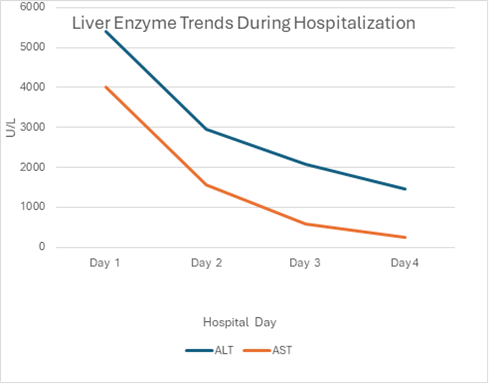
Figure 1: Aminotransferases declined rapidly within 72 hours.
Discussion
The patient’s initial presentation of diffuse abdominal pain and flank pain can be explained by splanchnic hemodynamic changes and renal hypoperfusion in the acute decompensated heart failure [6]. Such symptoms may complicate early diagnostic assessment since they mimic primary hepatobiliary or gastrointestinal conditions [7]. The rapid improvement in aminotransferases within 72 hours of aggressive diuresis and inotropic support strongly supports ischemic hepatopathy rather than primary hepatic pathology [8]. The transient rise in bilirubin with subsequent improvement further aligns with the expected biochemical course of hypoxic liver injury.
Massive aminotransferase elevation exceeding 5000 U/L narrows the differential diagnosis to a limited group of conditions, most notably ischemic hepatitis and drug-induced liver injury [9].
In this patient, the hepatocellular pattern evidenced by marked AST/ALT elevation with relatively normal alkaline phosphatase, associated coagulopathy, lactic acidosis, and acute kidney injury initially raised concern for severe intrinsic liver disease or toxin exposure. Acetaminophen toxicity was excluded by an undetectable level, and acute viral hepatitis was ruled out by negative serologies and absence of active HCV viremia. The rapid decline in transaminases without antiviral or immunosuppressive therapy argued against viral or autoimmune hepatitis.
Ischemic hepatopathy is a clinical syndrome characterized by compromised blood flow to the liver secondary to congestive heart failure, circulatory failure and septic shock [10,11]. In the setting of right heart failure, back up pressure leads to hepatic congestion, due to impaired hepatic venous outflow [12]. This will ultimately result in decreased hepatic blood flow, hypoxia, ischemic hepatitis, centrilobular necrosis [10,11].
In rare cases, where there is delay in the controlling the primary cause fibrosis, cirrhosis and hepatocellular carcinoma might ensue [13]. The clinical criteria that fulfill the diagnosis of congestive hepatopathy includes rapid and sustained elevation of plasma transaminases in the patient with heart or circulatory failure where all other causes of hepatic injury including viruses, drugs, and toxins are excluded [11]. By case definition, a 10-fold elevation of aminotransferases is necessary for the diagnosis of ischemic hepatitis [2, 14].
In a systemic review and meta-analysis, it was revealed that ischemic hepatitis was found in 2.5 cases for every 100 ICU admissions and 2 cases out of all hospital admissions [2]. Ischemic hepatitis were noticed in 78% and 23% of the patients with heart failure and sepsis respectively [2]. In case-control cohort study, 31 patients with right sided heart failure and hepatic congestion developed ischemic hepatitis [20-fold elevation in aspartate aminotransferases] when they are exposed to severe hypotension as compared to control cohort who sustained non-hepatic trauma and associated low blood pressure [15].
Heart failure is the predominant risk factor for instigating the commencement of cellular events that spark off the onset of ischemic hepatitis [10,11,14,16,17]. That being said, right heart failure with subsequent elevated filling pressures will drive further chain of events that evoke decreased hepatic perfusion, oxygen saturation and nutrition supply to the hepatocytes, thus bringing to light ischemic hepatitis [16]. Over and above right heart failure, presence of supplementary risk factors such as septic shock, prolonged hypotension, hypovolemic shock, respiratory failure, cardiothoracic surgery, obstructive sleep apnea, acute myocardial infarction, pulmonary embolism, cardiac tamponade, extensive burns and anemia might lower the threshold for unfolding ischemic hepatitis [1,3,15,18-21]. In addition, other risk factors that heighten the propensity to emanate ischemic hepatitis include obesity, physical inactivity, hypertension, diabetes, coronary artery disease, and valvular heart disease [22].
Cardiac output is responsible for hepatic blood flow via hepatic arteries and portal venous system [11]. Central zone of hepatic lobule receives the most blood supply whereas peripheral zones received least blood supply [11]. Due to physiological hepatic autoregulation in place, heart failure induced increased portal blood flow causes low adenosine levels, thence ultimately leading to hepatic arteriolar vasoconstriction and reduced hepatic arterial blood flow [11, 23]. That being said, the histological changes in ischemic hepatitis can range from centrilobular dilation, peri-perivenular fibrosis, to hepatic atrophy [2].
The underlying pathological events that are instrumental in initiating, progressing and sustaining the downstream cellular events for culminating the onset of ischemic hepatitis include reduced hepatic perfusion, hepatic venous congestion, peri-sinusoidal edema, reduced oxygen & nutrition delivery to hepatocytes, reperfusion injury, and hepatocellular damage [1,11,16]. In addition, impaired hepatocullar function, fibrogenesis, structural changes, inflammatory mediators, hepatic ischemia, and necrosis can also contribute to its inception [10]. In the presence of co-existent sepsis, distributive shock, circulating endotoxins and inflammatory mediators can contribute to hepatic hypoxia due to increased oxygen requirements and impaired ability of hepatocytes to utilize oxygen [24]. Cellular hypoxia triggers chain of events including ATP depletion, decrease in energy dependent pathways, increased anerobic glycolysis, increased lactate accumulation, potassium influx, increased calcium uptake, activation of proteases, & phospholipases, thus instigating cytoskeletal perturbation, membrane damage, cell lysis, apoptosis and necrosis [25].
In patients with heart failure, even 20 minutes of transient decrease in hepatic hypoperfusion can culminate in hepatic necrosis due to combination of downstream cellular events including inflammation, oxidative stress and overproduction of reactive oxygen species [15,25,26].
In a typical clinical scenario, an ICU patient with severe heart failure or chronic respiratory failure or sepsis presents with symptoms of right heat failure with abdominal pain, nausea, vomiting, fatigue, weakness, shortness of breath, leg swelling, ascites, and right upper quadrant pain [1,3,10,13,25,27]. Physical examination might reveal hepatomegaly, hepatojugular reflex and pedal edema [1,10]. Lab work up might reveal 10-20-fold elevation of transaminases, raised LDH levels and elevated bilirubin [1,28,29]. In a typical scenario, rapid and transient elevation of aminotransferases occurs within 24 h followed by transient resolution within next 1-3 days and complete normalization within next 2-3 weeks is hallmark of ischemic hepatitis [3, 20]. The rise in aminotransferases might signal the presence of hepatic venous congestion [30]. Ascertaining the cardiac source in ischemic hepatitis can be performed by measuring Brain Natriuretic Peptide (BNP), which is typically elevated above 364 pg/ml [31]. Other non-specific findings that might indicate liver failure include increased ammonia levels reduced prothrombin levels, increased lactate, hyperglycemia and increased INR [1,17,32,33].
Diagnosis of ischemic hepatitis is mostly dependent upon replying of classic clinical triad of rapid and transient elevation of transaminases in a critically ill patient admitted to ICU where other causes of hepatic injury are excluded (Viruses, Drugs, Toxins, Autoimmune, and trauma) [1]. Liver biopsy is rarely required to corroborate the diagnosis which might show centrilobular necrosis [1,34]. Bedside ultrasonography performed in a patient with ischemic hepatitis might reveal dilation of inferior vena cava and supra hepatic veins due to hepatic congestion, hepatomegaly, fatty liver, portal hypertension, splenomegaly or ascites [1,35]. CT scan/MRI are usually not usually necessary to confirm the diagnosis, but might be useful in excluding other diagnosis [1]. Some clinical studies had proposed the utility of indocyanine green (ICG) and hepatic veinous pressure monitoring for assessing the changes in the hepatic blood flow for diagnostic and prognostic purposed[1,36,37]. Echocardiography might be needed to evaluation the heart function, and ejection fraction and valvular abnormalities [38].
The management of ischemic hepatitis entails management of root cause of hepatic congestion that is congestive heart failure along with regular monitoring of hepatic functions tests. The four pillars of Guideline Directed Medical Therapy (GDMT) including ACE [angiotensin‐converting enzyme inhibitors/ARBs [angiotensin receptor blockers]/ARNI [angiotensin receptor-neprilysin inhibitor], BB [Beta-blockers], MRA [Mineralocorticoid receptor inhibitor], SGLT2i [sodium-glucose cotransporter-2 inhibitors,] needs to utilized for efficient management for heart failure [39]. In low cardiac output states or cardiogenic shock, resorting to usage of inotropes such as dobutamine can be very beneficial for augmenting cardiac output and improving splanchnic blood flow to the liver tissues [40,41].
Few clinical trials have shown that N-acetyl cysteine (NAC) would be beneficial in improving clinical outcomes and survival mortality in non-acetaminophen related liver failure [42,43]. In case-report by Parvathaneni et al, administration of NAC in a patient with shock liver secondary to sepsis resulted in modest improvement in clinical outcomes [44]. In end stage heart failure with low ejection fraction, usage of Left Ventricular Assist Devices (LAVD), and cardiac resynchronization therapy as well as heart transplantation has been shown to be beneficial in improving quality of life and survival [45-47].
Continuous monitoring of liver enzymes including aminotransferases, bilirubin, alkaline phosphatase, gamma-glutamyl transferase, and albumin, along with scores like the Model for End-Stage Liver Disease (MELD) to ensure recovery of liver function is very much mandatory in this clinical scenario [2,48].
Conclusion
The pivotal finding was newly diagnosed severe biventricular systolic dysfunction with evidence of elevated right-sided pressures. Hypoxic hepatitis is most associated with cardiac failure and may occur even in the absence of documented hypotension. In this setting, reduced forward cardiac output combined with hepatic venous congestion likely resulted in impaired hepatic oxygen delivery. While this patient exhibited signs of volume overload including pleural effusions, reduced right ventricular function, and a dilated non-collapsing inferior vena cava, the homogenous echotexture from liver ultrasound supports acute cardiogenic liver injury rather than acute decompensation superimposed on established chronic congestive hepatopathy [49].
Take home messages
- ·Ischemic hepatopathy is a clinical syndrome that transpires when there is low blood flow in combination with hepatic congestion.
- ·All the causes of hepatocellular damage including drugs, viral, toxins and autoimmune needs to be excluded.
- ·The most common cause is congestive heart failure, although causes including respiratory failure, sepsis, severe hypotension, severe pulmonary hypertension, cardiomyopathies and constrictive pericarditis are also documented in literature.
- ·The underlying pathophysiology entails reduced hepatic perfusion, hepatic venous congestion, peri-sinusoidal edema, reduced oxygen & nutrition delivery to hepatocytes, reperfusion injury, and hepatocellular damage.
- ·In a typical clinical scenario, ICU patient with heart failure presents with abdominal pain, jaundice, ascites, weakness, nausea, vomiting and pedal edema with elevation of liver enzymes.
- ·Lab workup might reveal A 10–20-fold raise in aminotransferases which is might partially resolve in next 1-3 days.
- ·Monitoring heart function with BNP, echocardiography might be essential.
- ·The main therapy entails efficient management of heart failure with GDMT as well as inotropes.
- ·In few studies, NAC has been shown to be beneficial by alleviating hepatic filling pressure
- ·Most cases resolve spontaneously with normalization of liver enzymes with recuperation of heart function and restoration of liver blood flow
- ·In rare cases where acute liver failure ensues, reassortment to advanced treatment strategies including LAVD, CRT and heart transplantation has shown to improvement in liver function, clinical outcomes and mortality in ischemic hepatitis
Conclusion
This case underscores the importance of early cardiac evaluation in patients presenting with extreme transaminase elevation, particularly when accompanied by abdominal symptoms and signs of volume overload. Distinguishing ischemic hepatitis from other causes of severe hepatocellular injury is critical, as management depends on prompt correction of the underlying hemodynamic disturbance rather than liver-directed therapy. It also highlights the highly reversible nature of ischemic hepatopathy when the underlying disturbance is promptly addressed.
References
- Waseem N, Chen PH. (2016). Hypoxic Hepatitis: A Review and Clinical Update. J Clin Transl Hepatol. 4: 263-268.
- Tapper EB, Sengupta N, Bonder A. (2015). The Incidence and Outcomes of Ischemic Hepatitis: A Systematic Review with Meta-analysis. Am J Med. 128: 1314-21.
- Henrion J. (2012). Hypoxic hepatitis. Liver Int. 32: 1039-52.
- Henrion J, (2000). Ischemia/reperfusion injury of the liver: pathophysiologic hypotheses and potential relevance to human hypoxic hepatitis. Acta Gastroenterol Belg. 63: 336-47.
- Hirao H, Nakamura K, Kupiec-Weglinski JW. (2022). Liver ischaemia-reperfusion injury: a new understanding of the role of innate immunity. Nat Rev Gastroenterol Hepatol. 19: 239-256.
- Sundaram V, Fang JC. (2016). Gastrointestinal and Liver Issues in Heart Failure. Circulation. 133:1696-703.
- Samsky MD, Patel CB, Dewald TA, Smith AD, Felker GM, et al. (2013). Cardiohepatic interactions in heart failure: an overview and clinical implications. J Am Coll Cardiol. 61: 2397-2405.
- Ebert EC. (2006). Hypoxic liver injury. Mayo Clin Proc. 81: 1232-6.
- Mahl TC. (1998). Approach to the patient with abnormal liver tests. Lippincotts Prim Care Pract. 2: 379-89.
- Haddadin R, Aboujaoude C, Trad G. (2024). Congestive Hepatopathy: A Review of the Literature. Cureus. 16: e58766.
- Ciobanu AO, Gherasim L. (2018). Ischemic Hepatitis - Intercorrelated Pathology. Maedica (Bucur). 13: 5-11.
- Hilscher M, Sanchez W. (2016). Congestive hepatopathy. Clin Liver Dis (Hoboken). 8: 68-71.
- Myers RP, Cerini R, Sayegh R, Moreau R, Degott C, et al. (2003). Cardiac hepatopathy: clinical, hemodynamic, and histologic characteristics and correlations. Hepatology. 37: 393-400.
- Lightsey JM, Rockey DC. (2017). Current concepts in ischemic hepatitis. Curr Opin Gastroenterol. 33: 158-163.
- Seeto RK, Fenn B, Rockey DC. (2000). Ischemic hepatitis: clinical presentation and pathogenesis. Am J Med. 109: 109-13.
- McClairen C, Doering T. (2022). Case of Ischemic Hepatitis in the Setting of HFpEF Exacerbated by Persistent Atrial Fibrillation. J Community Hosp Intern Med Perspect. 12: 73-74.
- Henrion J, Schapira M, Luwaert R, Colin L, Delannoy A, et al. (2003). Hypoxic hepatitis: clinical and hemodynamic study in 142 consecutive cases. Medicine (Baltimore). 82: 392-406.
- Fuhrmann V, Kneidinger N, Herkner H, Heinz G, Nikfardjam M, et al. (2009). Hypoxic hepatitis: underlying conditions and risk factors for mortality in critically ill patients. Intensive Care Med. 35: 1397-405.
- Trilok G, Qing YC, Li-Jun X. (2012). Hypoxic hepatitis: a challenging diagnosis. Hepatol Int. 6: 663-9.
- Henrion J, MINETTE P, COLIN L, SCHAPIRA M, et al., (1997). Hypoxic hepatitis caused by severe hypoxemia from obstructive sleep apnea. J Clin Gastroenterol. 24: 245-9.
- Blich M, Edelstein S, Mansano R, Edoute Y. (2003). Ischemic hepatitis induced by severe anemia. Isr Med Assoc J. 5: 208-9.
- He J, Ogden LG, Bazzano LA, Vupputuri S, Loria C, et al. (2001). Risk factors for congestive heart failure in US men and women: NHANES I epidemiologic follow-up study. Arch Intern Med. 161: 996-1002.
- Lautt WW. (1985). Mechanism and role of intrinsic regulation of hepatic arterial blood flow: hepatic arterial buffer response. Am J Physiol. 249: G549-56.
- Nelson DP, Samsel RW, Wood LD, Schumackeret PT. (1988). Pathological supply dependence of systemic and intestinal O2 uptake during endotoxemia. J Appl Physiol (1985). 64: 2410-9.
- Fuhrmann V, Jager B, Zubkova A, Drolz A. (2010). Hypoxic hepatitis - epidemiology, pathophysiology and clinical management. Wien Klin Wochenschr. 122: 129-39.
- Massip-Salcedo M, Roselló-Catafau J, Prieto J, Avíla MA, Peralta C. (2007). The response of the hepatocyte to ischemia. Liver Int. 27: 6-16.
- Fortea JI, Puente A, Cuadrado A, Huelin P, Pellon R, et al. (2020). Congestive Hepatopathy. Int J Mol Sci. 21: 9420.
- Cassidy WM, Reynolds TB. (1994). Serum lactic dehydrogenase in the differential diagnosis of acute hepatocellular injury. J Clin Gastroenterol. 19: 118-21.
- Jäger B, Dorlz A, Michl B, Schellongowski P, Bojic A, et al. (2012). Jaundice increases the rate of complications and one-year mortality in patients with hypoxic hepatitis. Hepatology. 56: 2297-304.
- Ford RM, Book W, Spivey JR. (2015). Liver disease related to the heart. Transplant Rev (Orlando). 29:33-7.
- Sheer TA, Joo E, Runyon BA. (2010). Usefulness of serum N-terminal-ProBNP in distinguishing ascites due to cirrhosis from ascites due to heart failure. J Clin Gastroenterol. 44: e23-6.
- Stravitz RT, Kramer AH, Davern T, Shaikh AOS, Caldwell SH, et al. (2007). Intensive care of patients with acute liver failure: recommendations of the U.S. Acute Liver Failure Study Group. Crit Care Med. 35: 2498-508.
- Birrer R, Takuda Y, Takara T. (2007). Hypoxic hepatopathy: pathophysiology and prognosis. Intern Med. 46: 1063-70.
- Henrion J, Schapira M, Heller FR. (1996). Ischemic hepatitis: the need for precise criteria. J Clin Gastroenterol. 23: 305.
- Denis C, De Kerguennecet C, Bernuau J, Beauvais F, Solalal AC. (2004). Acute hypoxic hepatitis ('liver shock'): still a frequently overlooked cardiological diagnosis. Eur J Heart Fail. 6: 561-5.
- Horvatits T, Kneidinger N, Drolz A, Roedl K, Rutter K, et al. (2015). Prognostic impact of ICG-PDR in patients with hypoxic hepatitis. Ann Intensive Care. 5: 47.
- Halle BM, Poulsen TD, Pedersen HP. (2014). Indocyanine green plasma disappearance rate as dynamic liver function test in critically ill patients. Acta Anaesthesiol Scand. 58: 1214-9.
- Marwick TH. (2015). The Role of Echocardiography in Heart Failure. Journal of Nuclear Medicine. 56: 31S-38S.
- Berry NC, Sheu YS, Chesbrough K, Bishop RC, Vupputuri S. (2025). Guideline-directed medical therapy rates in heart failure patients with reduced ejection fraction in a diverse cohort. ESC Heart Fail. 12: 1861-1871.
- El Mokhtari, N.E., et al., Inotropic therapy for cardiac low output syndrome: comparison of hemodynamic effects of dopamine/dobutamine versus dopamine/dopexamine. Eur J Med Res, 2007. 12(11): p. 563-7.
- Kram HB, Evans T, Bundage B, Shoemaker WC. (1988). Use of dobutamine for treatment of shock liver syndrome. Crit Care Med. 16: 644-5.
- Nabi T, Nabi S, Rafiq N, Shah A. (2017). Role of N-acetylcysteine treatment in non-acetaminophen-induced acute liver failure: A prospective study. Saudi J Gastroenterol. 23: p169-175.
- Lee WM, Hynan LS, Rossaro L, Fontana RJ, Stravitz RD, et al., (2009). Intravenous N-acetylcysteine improves transplant-free survival in early stage non-acetaminophen acute liver failure. Gastroenterology. 137: 856-64, 864.e1.
- Parvataneni S, Vemuri-Reddy S. (2020). N-acetyl Cysteine Use in the Treatment of Shock Liver. Cureus. 12: e7149.
- Birati EY, Jessup M. (2015). Left Ventricular Assist Devices in the Management of Heart Failure. Card Fail Rev. 1: 25-30.
- Long T, Yu Y, Cheng S, Huang H, Hua W. (2025). Prognostic value of the modified Model for End-Stage Liver Disease score in patients treated with cardiac resynchronization therapy. Heart Rhythm O2. 6: 339-349.
- Chokshi A, Cheema FH, Schaefle KJ, Jiang J, Collado E, et al., (2012). Hepatic dysfunction and survival after orthotopic heart transplantation: application of the MELD scoring system for outcome prediction. J Heart Lung Transplant. 31: 591-600.
- Yancy CW, Jessup M, Bozkurt B, Butler J, Drazner MH, et al. (2013). 2013 ACCF/AHA guideline for the management of heart failure: a report of the American College of Cardiology Foundation/American Heart Association Task Force on practice guidelines. Circulation. 128: e240-327.
- Xanthopoulos A, Starling RC, Kitai T, Triposkiadis F. (2019). Heart Failure and Liver Disease: Cardiohepatic Interactions. JACC Heart Fail. 7: 87-97.