Samir Akram Sakka*
Department of Orthopaedic Surgery, Al Balqa Applied University, Al Balqa, Jordan
*Corresponding author: Samir Akram Sakka, Deparment of Orthopaedic Surgery, Al Balqa Applied University, Al Balqa, Jordan
Received Date: 15 October 2026
Accepted Date: 22 October 2026
Published Date: 30 October 2026
Citation: Sakka S (2026) Rising Needle Sign During Lumbar Facet Injection Technique: A Useful Sign in Helping to Localise the Facet Joint and Reducing Radiation Exposure. Ann Case Report 11: 2677. DOI: https://doi.org/10.29011/2574-7754.102677
Abstract
Introduction: Back pain is a common problem affecting the general population. Conservative treatment is the main treatment method. Facet injection is an integral part of this conservative treatment. Lumbar facet injection can be carried out under florescent radiography, CT Scan guided or recently under ultrasound. Several attempts are often needed to achieve the localising required by the operator prior to injection of facet whether using the intra-articular or the peri-articular technique. This meant that even in the expert hands increased radiation exposure to both the patient and the operator. In the case when ultrasound is used there is an increase in time and pain or discomfort to the patient with every additional localising attempt. This is more evident for new operators and trainees.
Aim: The aim the study is demonstrate the rising needle sign during facet injection its anatomical basis and its usefulness during facet injection and reducing radiation by helping to localise the facet quickly and thus reducing radiation time.
Method: Having observed the rising needle during the localisation of the facet on the AP view, we set to confirm this by studying the anatomical models and surgical anatomy to explain and demonstrate the sign.
Conclusion: After detailed anatomical consideration the author used the sign routinely during the procedure and observed significant marked reduction of the time usage of the Image intensifier exposure when more than one facet is injected and using two needles simultaneously helping the operator to reduce the number of attempts in localising the needle for optimal injection, the time needed to do so, and the radiation exposure to the patient and the operator.
Keywords: Facet injection; Fluoroscopy; Radiation; Rising Needle sign; Spine.
Introduction
Back pain is a common problem affecting the general population. Conservative treatment is the main method of treatment. Facet injection is an integral part of this conservative treatment. Lumbar facet injection can be carried out under florescent radiography, CT scan guided or recently under ultrasound [1-4]. Several attempts often needed to achieve the localising required by the operator prior to injection whether using the intra-articular or the peri-articular technique. This meant even in the expert hand increased radiation exposure to both patients and operators [5]. In the case when ultrasound is used there is an increase in time and pain or discomfort to the patient with every additional localising attempt. This is more evident for new operators and trainees, hence training using simulator had by used to reduce the radiation exposure.[6,7,8]. Facet injection under florescent radiography is the commonly used method [9]. We observed the rising of the needle when localising the facet as it reaches the desired position. To verify this we looked at the anatomy of the facet and its surrounding structures and its effects on the needle during its positioning. We then looked at the surgical anatomy during our open spinal procedures. Subsequently using this observational clinical signed helped to reduce the rotation time significantly. We describe the Rinsing Needle Sign, which help the operator to reduce the number of attempts in localising the facet, the time needed to do so, and the radiation exposure to the patient and the operator. We recommend observing and utilising this sign by expert operators and new trainees alike. This article was previously presented as a meeting abstract at the 2021 Global Spine Congress on 3-6 November 2021.
Methods
Keywords: Facet joint pain contributes significantly to lower back pain. Image intensifier x-ray guidance is used to locate the facet joints. Radiologically there are two recognized approaches for facet injection; the ‘scotty dog’ and the AP views with no significant difference between the two views approaches [9]. There are also two methods described for delivering the injections to treat facet joint pain; intra-articular and peri-articular with no significant difference of pain relief outcome between them [10]. We use the AP view utilising method on the image intensifier and the peri-articular approach for delivering the injectate. With regards to the visualisation of the facet joint itself, the target point will be the midpoint of the silhouette of the joint cavity. If this is not visualised, it could be either due to the destructive arthritic process or mal position of the x-ray beam [9]. Having observed the rising needle during the localisation of the facet on the AP view, we set to confirm this by studying the anatomical models and surgical anatomy to explain and demonstrate the sign. After detailed anatomical consideration the author used the sign routinely during the procedure and observed significant marked reduction of the time usage of the Image intensifier exposure when more than one facet is injected and using two needles simultaneously.
Discussion
The spine is the most common source of chronic pain and the second most common reason for a patient to consult a physician [1,2]. Back pain is also a common problem that afflicts 70%-85% of people in their lifetime and 15%-45% annually [1]
About two-thirds of the population suffers from back pain at some point of time during their life span and this symptom incapacitates 20% of them for long periods (>4 weeks) [2,3].
Conservative treatment of degenerative lumbar spine is the main steam and the common initial treatment. Facets injections is part of this conservative treatment. Fluoroscopy is an important part of interventional pain management procedures in modern medical practices. The purpose of fluoroscopy or other radiological modality CT/ US in interventional pain management is to place the needle specificity and accurately to deliver the injectate. When facet injection is performed under fluoroscopic visualisation, it is accurate and clinically useful in the diagnosis and therapeutic management of chronic spinal pain. [11,12].
The amount of radiation used depends on the mode used ie CT scan or Image intensifier. The former is more radiation risk to the patient and the operator [13]. Interventional procedures are associated with high radiation doses for both patients and surgeons, who often have to put their hands in the beams of the fluoroscope. The technique using fluoroscopy remains the mainstream. The experience and the seniority of the operator are recognised factors as it has been shown that trainee uses more X-ray radiations [7, 9].
The use of fluoroscopy-guided procedures has increased dramatically over the years. The dose arising from these procedures is estimated to be about 10% of the total medical exposure [6]
Physicians who used fluoroscopy should be aware of radiation exposure and make every effort to minimize radiation dose. Most doctors are not aware of their level of exposure to radiation [5-8,].
To reduce the risk from ionizing radiation, it is essential to minimize radiation dose. Monitoring, as well as the development of new interventional techniques possibilities like our proposed one [5].
Learning curve of radiology residents during training in fluoroscopy-guided facet joint injections has been shown to improve using simulation-based training programme [6-8].
We describe the anatomical basis for the RISING Needle Sign and illustration in the lumbar facet and illustrate it accordingly. Looking at the bony anatomy of the lumbar facet within the spinal segment from the caudal to the cranial direction could be easily seen that the facet is the highest point forming a prominent nodular structure between the lamina above and the lamina below (Figure 1). In surgical anatomy, the facet height is increased by the capsule. Therefore as the injection needle is inserted and moved along from the lamina above or below towards the facet it rises. The maximum rise is when it reaches the top of the middle of the facet in the cranially-caudally direction (Figure.1).
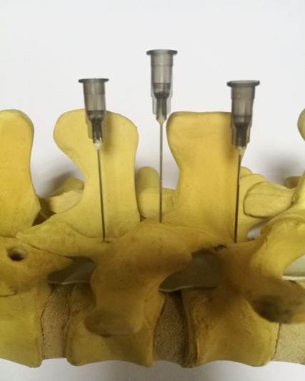
Figure 1: Lateral facet view of spine model showing the dome shaped facet rising higher than the adjacent bony landmarks caudally and cranially to it. The ‘rising needle’ is demonstrated.
Moving from lateral to medial ie from the transverse process towards the spinous process in the midline, the spinal needle will rise over the facet then down before it rises again when it reaches the midline over the spinous process (Figure. 2). The amount by which the needle can rises is predictable from the size face been measured in the normal patients without facet hypertrophy by Panjabi to be 18.4 mm. From the rising needle prospective about 2 cm maximum vertical needle movement or difference [14].
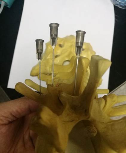
Figure 2: Transverse view of a spinal model with needles demonstrates the ‘rising needle sign’ as the needles are placed medially on the lamina and laterally on transverse process and on the dome of the facet in between.
Placing the needle in the mid line over the spinous process or near it will give a falsely positive impression of a rising needle. In the real and pathological anatomy the increase in the height of faces or the rise of the needle, is more noticeable because of facet hypertrophy and the osteophytes formation. This is illustrated in a patient prior and post exposed during surgery with the placement of the needle demonstrating the Rising Needle Sign (Figures 3 and 4).
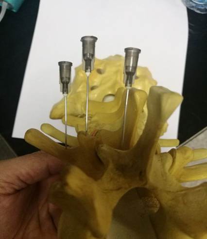
Figure 3: The Rising Needle sign on the Rt with the needle on the top of the facet while the left the needle placement on the pedicle at a lower level with near 2 cm difference in height.
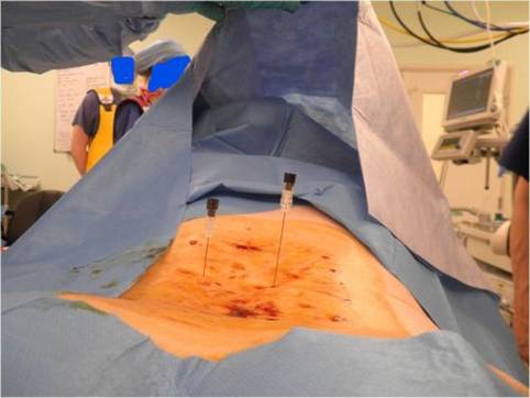
Figure 4: Intra-operative surgical anatomy demonstration of the ‘Rising needle’ ; on the right facet ( rising) and left needle on the pedicle below the facet with needle sinking lower than the one on the right.
This came about as observation while carrying the procedure using double needle by an experienced spine surgeon {author}who is familiar with the topographic anatomy and the bone landmark of the spine. As facets especially in the lumbar region form a bone prominence rises above the level of the adjacent anatomical structures such as the lamina superiorly and inferiorly and the transverse process laterally. Therefore, as the needle is inserted at the lever required under fluoroscopy until it reaches in depth the desired bony landmark mark of the expected facet then it will rise highest above the skin surface when it is on the middle of the facet. Therefore, the operator can use this to maximize the position of the needle and after it rises most, then he can use the Image intensified to verify the final position prior to deliver the injectate thus reducing the operating time and reducing repeated radiation exposure. This can also be of help using ultrasound guided needle procedures.
Conclusion
The Rising Needle Sign is a very useful sign in helping localising the facet during needle placement. In our experience this has reduced the exposure time and procedure time significantly. This will also be especially useful for novices and infrequent users helping them in reducing radiation exposure and the operation time.
Declaration
There is no conflict of interest or financial interest to declare
References
- Andersson GB. (1999). Epidemiological features of chronic low-back pain. Lancet. 354: 581-585.
- Hellsing A, Bryngelsson I. (2000). Predictors of musculoskeletal pain in men. A twenty year follow-up from examination at enlistment. Spine. 25: 3080-3086.
- Deyo RA, Weinstein JN. (2001). Low back pain. N Engl J Med. 344: 363-370.
- Wu T, Zhao WH, Dong Y, Song HX, Li JH. (2016). Effectiveness of Ultrasound-Guided Versus Fluoroscopy or Computed Tomography Scanning Guidance in Lumbar Facet Joint Injections in Adults With Facet Joint Syndrome: A Meta-Analysis of Controlled Trials Arch Phys Med Rehabil. 97: 1558-1563.
- Manchikanti L, Cash KA, Moss TL, Pampati V. (2002). Radiation exposure to the physician in interventional pain management Pain Physician. 5: 385-93.
- Proschek D, Kafchitsas K, Rauschmann MA, Kurth AA, Vogl TJ, et al. (2008). Florian Geiger Reduction of radiation dose during facet joint injection using the new image guidance system SabreSource™: a prospective study in 60 patients. Eur Spine J. 18: 546-553.
- Dias TR, Costa Alves Junior JDDD, Abdala A. (2017). Learning curve of radiology residents during training in fluoroscopy-guided facet joint injections Radiol Bras. 50: 162-169.
- Faulkner AR, Bourgeois AC, Bradley YC, Hudson KB, Heidel RE, et al. (2015). Simulation-based Educational Curriculum for Fluoroscopically Guided Lumbar Puncture Improves Operator Confidence and Reduces Patient Dose. Acad Radiol. 22: 668-73.
- Al-Tawil K, Lopez D, Blackman M, Suresh S. (2018). Oblique “Scotty dog” versus antero-posterior (AP) views in performing x-ray guided facet joint injections J Clin Orthop Trauma. 9: S145-S148.
- Kershen LM, Nacey NC, Patrie JT, Fox MG. (2018). Fluoroscopically Guided Facet Injections: Comparison of Intra-Articular and Periarticular Steroid and Anesthetic Injection on Immediate and Short-Term Pain Relief. AJNR Am J Neuroradiol. 39: 2161-2165.
- Peh WCG (2011). Image-guided facet joint injection Biomed Imaging Interv J. 7: e4.
- Sehgal N, Shah RV, McKenzie-Brown AM, Everett CR. (2005). Diagnostic utility of facet (zygapophysial) joint injections in chronic spinal pain: a systematic review of evidence. Pain Physician. 8: 211-224.
- Maino P, Presilla S, Colli Franzone PA, Van Kuijk SMJ, Perez RSGM, et al. (2018). Radiation Dose Exposure for Lumbar Transforaminal Epidural Steroid Injections and Facet Joint Blocks Under CT vs. Fluoroscopic Guidance. Pain Pract. 18: 798-804.
- Panjabi MM, Oxland T, Takata K, Goel V, Duranceau J, et al. (1993). Articular facets of the human spine. Quantitative three-dimensional anatomy Spine. 18: 1298-310.